Cindy Tung / Dr. Susan Gauthier - Week 4
This week, I shadowed Dr. Gauthier for the first time in her outpatient clinic as she saw her MS patients. MS results from the body's immune system attacking myelin sheaths surrounding the nerve fibers. Because there is no cure, patients visit the physician annually or bi-annually to track treatment plans and monitor disease symptoms. Most of the patients we saw have been seeing Dr. Gauthier for close or more than 10 years. During the visit, Dr. Gauthier pulls up a side-by-side view of their most recent MRI vs from a year ago to review any lesions in the brain or spinal cord. Physical examinations such as walking speed, hand-eye coordination, bladder function, and limb reflexes are also conducted to evaluate the dosage of drugs prescribed for the patients. While most of the patients had little to no symptoms, some patients will digress to progressive MS, which causes symptoms to gradually get worse, usually resulting in an inability to walk.
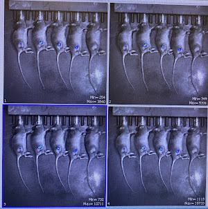
The tumor-injected mice from last week were checked up on every day to ensure they are not sick or have oversized tumors. This week, we injected them with luciferin in order to see the tumor in a IVIS in vivo CT imaging system. Under sedation, images are taken at 5, 10, 15, and 20 minutes after injection to dictate where there is most fluorescence in the tumors. The mice are then weighed to track the effect of the tumor on their body weight.
Time-Stamp Images Chamber Set-Up
For my research, I've been working with students in the lab to understand image post-processing for source separation in brain MRI scans. While the process is complex, I've been breaking down the steps from background filtering, unwrapping phases, convolution/deconvolution, etc. I've been given some small data sets and plan to practice performing analysis in the coming weeks.


Comments
Post a Comment