Cindy Tung / Dr. Susan Gauthier - Week 6
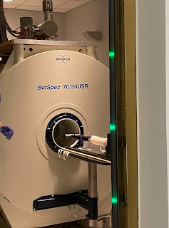
This week, we imaged our mice that were injected with breast cancer cells from last week. We measured their tumor sizes and put them in a Bruker 7T MRI for small animal imaging. A method called intravenous tail vein injection is used for inducing gadolinium. Gadolinium was used for image contrast, IV for hydration, and cyclophosphamide for chemotherapy.
Additionally, we performed another round of cell culturing on the SCC VII (head and neck) cancer cell line. The cell growth plates are first derived from frozen cells (seeding phase), followed by a passage phase where cells continue to proliferate. It is important that no bubbles are introduced during the plating stage as cells will not grow in those areas.
SCC VII Cell Line under Microscope
For the clinic this week, I was able to observe an ACL and total knee repair by Dr. Robert Marx at HSS. Additionally, I also saw an aortic valve replacement & ventricular septal defect repair in the cardiology department. It was amazing to see different teams (anesthesiology, perfusionists, surgeons, nurses, surgical technicians) in the operating room work together to provide the best care for the patient. The preparation for the surgery alone can take up to two hours, setting up sterilization, gathering the appropriate tools/machines, and cross-checking the procedure.


Comments
Post a Comment